Pleuronectiformes: M I A4
Unknown
|
Egg diameter in µm |
Number of oil globules |
Diameter of oil globule in µm |
Yolk texture |
Perivitelline space |
Position of oil globule at hatch |
Gut length at eye- pigment stage |
Myomeres |
|
1610 |
multiple |
NA |
clear |
narrow |
stern |
50% of NL |
30+ |
.
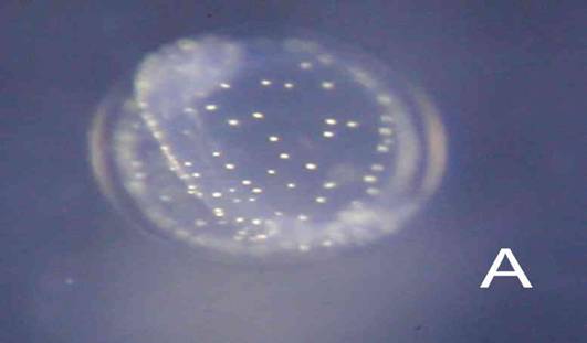
Egg: The egg has only been seen twice (A), and on the first occasion was mistaken for MIIA1. The late embryo and yolksac are lightly covered in white pigment spots (A). Incubation is uncertain; probably about 3 days.
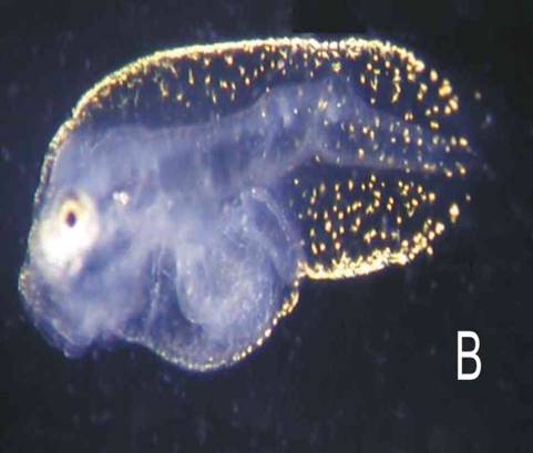
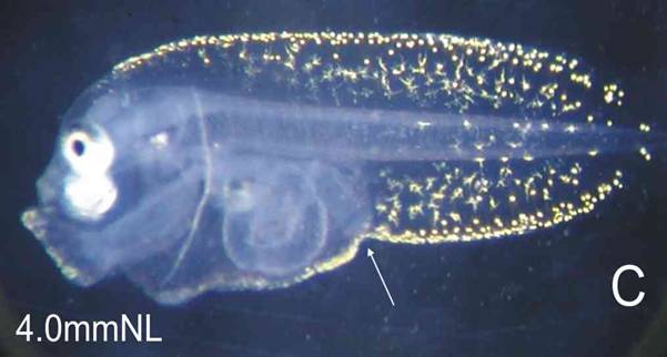
Larva: The 3-day larva has its finfolds covered in fine yellow spots (D), consolidated along the dorsal and ventral edges, to form a dense trim (B). The 5-day larva is much the same, but the right eye is already moving, suggesting this is a pleuronectid or soleid. B: 3 days, C: 5 days (25°C). The larva in Plate B has a distorted notochord.
Judging from the rapid onset of eye migration, this is probably one of the smaller soles.
The egg has only been recognised on three occasions, in February and October 2004, and April 2007. No larvae have yet been sequenced for a DNA barcode.